Magnetic Resonance Imaging (MRI)
Having a scan with us
At Bedford we are proud of our imaging team. We aim to provide the very best for our patients and visitors. Currently, the department has three state-of-the-art scanners located in South Wing.
At Bedford we are proud of our imaging team. We aim to provide the very best for our patients and visitors. Currently, the department has three state-of-the-art scanners located in South Wing.
Our facility uses the latest Siemens and GE scanners which provide high resolution images with minimal noise.
We understand that undergoing an MRI scan can be a daunting experience. We provide a calm and comfortable environment, with experienced Radiographers who will guide you through the entire process. All of our scanners are wide bore but if you do suffer with claustrophobia please call us and we can try to arrange for you to go into our short bore. We provide headphones so that you can listen to music whilst the scan is being performed to make the experience more relaxing and comforting to you.
Please ensure that you arrive at least 15 minutes before your appointment at MRI department, so that no inconvenience is caused to yourself or other patients.
The machine takes a few minutes to capture each image, so it is important to lie as still as possible throughout the scan. You will be provided with headphones or ear plugs to protect your hearing during your scan. Music will be provided through your headphones to listen to during the scan and to help you relax. Some scans you will be asked to do some breathing instructions, the Radiographers will speak with you before scan starts and will instruct you when the scan is happening to do the breathing exercises.
Very occasionally people can become too warm during an MRI scan, this is the main reason our MRI rooms are maintained at a low temperature and the MRI scanners have ventilation.
For some MRI scans it will be necessary for the Radiographer to give you an injection of contrast media into a vein, through a cannula during your scan. This is a clear, colourless dye that is used to make the images clearer during an MRI scan and help with diagnosis.
The contrast is a Gadolinium-based contrast agents (also known by their brand names Gadovist® Prohance® (Gadoteridol) and Primovist® (Gadoxetic disodium), which may be used during MRI scans. If you have any questions or concerns, please speak to your doctor or nurse. Further information about the contrast agent is available in the manufacturer’s patient information leaflet.
The contrast is not suitable for everybody. The radiographer or radiologist will decide if it is appropriate for you.
You must tell the radiographer or radiologist before your scan if you have any history of poor renal function or kidney problems, or if you have ever had an allergic reaction to a contrast agent.
The contrast does not usually cause any reaction. Some of the more common reactions include:
The reaction usually occurs during the injection or within the first hours afterwards; however, some can occur up to several hours later.
If you have any concerns about side effects, please speak to the staff caring for you.
A small amount of the contrast agent is excreted into the breast milk. There is no proven risk to the infant, but if you are worried you can discard the breast milk for 24 hours after the injection.
Once the MRI scan is complete, our Radiologists will interpret the images and prepare a report to send to your referrer.
It is important to follow-up with your healthcare provider to discuss your MRI results and discuss any next steps.
Brain MRI: MRI (Magnetic Resonance Imaging) is a valuable diagnostic tool used to assess the brain for a variety of abnormalities. It provides detailed images of the brain and its structures, helping healthcare providers identify potential causes for symptoms like headaches and other neurological issues. Overall, MRI provides clear, detailed imaging, making it a vital tool for evaluating a wide range of neurological conditions and guiding appropriate treatment.

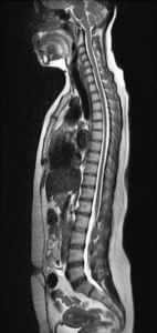
Spine MRI: A spine MRI (Magnetic Resonance Imaging) is commonly used to evaluate a range of spine-related issues. It provides detailed images of the spinal cord, vertebrae, discs, nerves, and soft tissues, helping doctors diagnose various conditions that may cause pain, discomfort, or neurological symptoms.
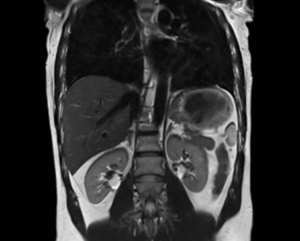
Abdominal MRI: An abdominal MRI (Magnetic Resonance Imaging) is a valuable diagnostic tool used to examine the organs and structures within the abdomen for various potential issues. It provides detailed images of soft tissues, which can help doctors identify a wide range of conditions. An abdominal MRI is a non-invasive imaging technique that provides detailed insights into various organs and structures within the abdomen. It helps healthcare providers diagnose and monitor a wide range of conditions, from benign to life-threatening issues, guiding treatment decisions and improving patient outcomes.
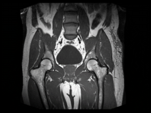
Pelvic MRI: pelvic MRI (Magnetic Resonance Imaging) is a highly effective diagnostic tool used to evaluate the organs and structures within the pelvic region. It provides detailed imaging of soft tissues, making it ideal for detecting a variety of conditions that can affect the pelvic area. a pelvic MRI is a versatile and non-invasive imaging tool that provides valuable information for diagnosing a wide variety of conditions in the pelvic area. It is particularly useful for assessing soft tissue abnormalities and guiding appropriate treatment decisions.
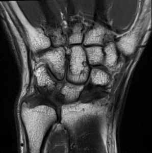
Musculoskeletal System: MSK (Musculoskeletal) examinations are clinical assessments used to evaluate the health and function of the musculoskeletal system, which includes bones, muscles, joints, ligaments, tendons, and other soft tissues. While MSK exams can involve any part of the body, they are commonly focused on extremities—such as the arms, hands, legs, and feet—to assess any issues that may affect movement, strength, or pain in these areas.
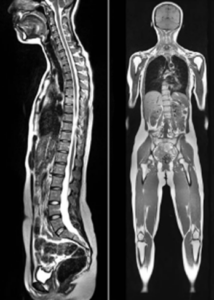
A whole-body MRI is a powerful diagnostic tool for obtaining comprehensive images of the entire body, helping detect a wide range of conditions. While it can take a long time and requires the patient to remain still, it is a non-invasive procedure with no radiation exposure, making it a safe option for assessing the overall health of the body.
Defecating Proctogram: This is a very specialized scan, which needs to be referred by a specific clinic and approved by a Senior Radiologist.
MRI is located via the Britannia Road entrance on 1st floor. You will need to go up the stairs and turn right and you will see a sign for MRI Suite 1, you need to report here for all MRI scans.
Getting to Bedford Hospital South Wing
There is a carpark on Britannia Road or Kempston Road. Parking charges apply unless you have a blue badge.
If you have any questions or concerns, or if you experience any of the symptoms listed above, please contact the MRI Department: